Published December 10, 2025 02:10PM
To MRI, Or Not to MRI? Experts Weigh in On The Pros and Cons of Medical Imaging
Some doctors believe the more info you have about a possible injury, the better; others say there are risks, especially early on in the treatment process. We spoke to experts on both sides of the issue.
(Photo: Canva Images)
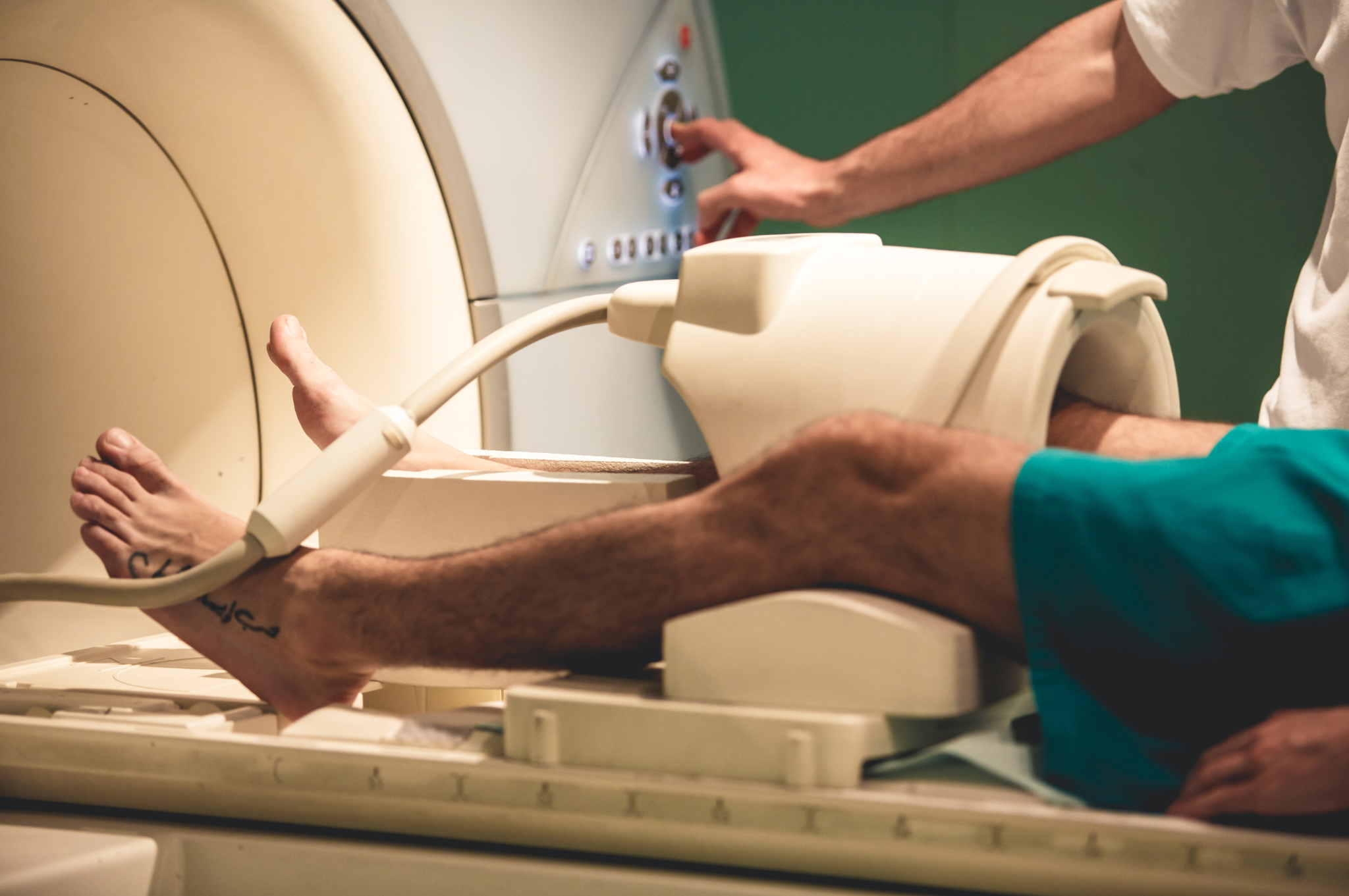
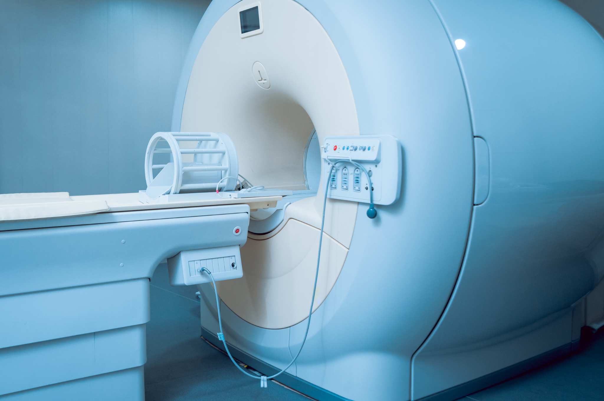
Many runners are familiar (read: too familiar) with joint pain and injuries. In fact, research suggests that every year, nearly half of all runners in the U.S. wrestle with some type of bodily ailment. When these issues arise and persist, a common response is to seek medical guidance. From there, many medical professionals recommend imaging, such as an MRI, X-ray, or ultrasound.
That approach may sound straightforward enough. After all, wouldn’t more information be a good thing, since it helps fine-tune a treatment plan? But it’s not always that simple, some healthcare experts say.
The Differing Views
Dan Ginader, P.T., D.P.T., a physical therapist in New York City, says that in certain cases, medical imaging results can have a psychological impact that’s more harmful than helpful. That’s because for active people over the age of 40 to 50, getting an MRI will likely reveal some sort of damage in the knees or back, even if they are completely pain-free and feeling healthy. This is due to natural age-related wear and tear that occurs in our bodies with time, he explains.
ADVERTISEMENT
Research shows simply learning you have damage is enough for your brain to start increasing pain or causing pain in that area, so for some people, the psychological damage of seeing imaging results can worsen their symptoms and make them feel like they’re “broken,” Ginader says.
That’s why, for mild and/or chronic pain, Ginader suggests runners try physical therapy first before jumping straight to imagery. Meanwhile, Kevin R. Stone, M.D., an orthopedic surgeon in San Francisco, takes a different approach. He believes that “whenever possible, you want to have good data about your injury.” An important part of that data-gathering process involves medical imaging. “In the 21st century, it makes no sense whatsoever not to have really good data to make good decisions on,” he says. “Just burying your head in the sand is almost never the right approach.”
To better understand the debate in the medical community about when and where imaging has its place in treating running-related pain, we spoke with three experts as well as a run coach who’s gotten several MRIs himself.
Roundtable Participants
Dan Ginader, P.T., D.P.T., is a physical therapist in New York City and author of The Pain-Free Body.
Kevin R. Stone, M.D., is an orthopedic surgeon at The Stone Clinic in San Francisco.
Joseph Frank, M.D., is an associate professor of internal medicine at University of Colorado Anschutz and co-director of the school’s Pain Science Program.
Will Baldwin is a distance runner and certified run coach in Denver. He’s gotten several MRIs in his nearly two-decade-long running career.
ADVERTISEMENT
The responses below reflect four separate conversations. Answers have been edited for length and clarity.
How did you arrive at your belief surrounding medical imaging?
Ginader: One of the major things taught in school, at least as it surrounds imaging, is the prevalence of damage that will show up, especially in people that are in their 40s and 50s. And the research shows that the prevalence of damage that shows up on imaging far outweighs symptoms that people feel.
Just because something looks damaged on imaging, it doesn’t necessarily mean that it is what’s leading to your symptoms because there are so many symptom-free people that show damage on an image. Also, I’ve been treating patients for close to a decade now, and I’ve seen the change in just how learning a result can change how they feel about their pain.
ADVERTISEMENT
What’s your take on Dr. Ginader’s stance?
Stone: The first thing that you need to know is that joints of the body can last a lifetime of sport and exercise, including running. They do not degrade unless they’re either injured or diseased, and so articular cartilage can last a lifetime.
The next part that’s important to understand is that if you’re having pain, pain is a good indicator of damage. Damage, when diagnosed early, is often much easier to treat and cure than if diagnosed late. Injury diagnosis is best made in the following ways: good patient history, good physical exam, high-quality X-ray, and high-quality MRI.
Can you elaborate on the disconnect between the damage that’s seen on an image and the actual symptoms people feel?
Ginader: The disconnect exists because pain is far more complex than mechanical damage. Pain is so deeply tied into your prior experiences, the context of the situation, even your current mood. Pain can greatly change based on how good of a night’s sleep you had, or based on your hormone levels, or based on a limitless amount of factors that are not just mechanical damage. And when you have a chronic condition, or you have a more minor condition, it makes that mechanical damage even a smaller piece of that puzzle. There’s research that shows that just the expectation of pain can cause pain. If you dive into pain science, there’s a really great TEDx Talk by Lorimer Moseley, an Australian physiotherapist, who spearheaded the popularization of pain science research.
Stone: This disconnect is why you always need a good combination of expert physical exam, history, X-ray, and MRI. They all go together, and when you have that combination together, then you can make a decision if something is seriously injured, is not seriously injured, needs repair, does not need repair, but it takes a combination. We see people with very small injuries—very small meniscus tears, for instance—that have debilitating pain. And we see other people with major injuries that have almost no pain whatsoever. And so understanding the individual and their exam determines a lot about whether or not something needs to be treated and whether or not it will go on to further damage their joints.
ADVERTISEMENT
What’s been your experience with MRIs?
Baldwin: I had two major incidents where an MRI actually completely changed what we did in the process. The most recent one was two years ago. I had a lot of pain around my calf and saw three different PTs. Everyone was like, “Yeah, this is like a mild calf strain.” So I did what you do for a mild calf strain: I changed the amount of load placed on it and did a lot of PT. But it just got worse and worse and worse. Eventually, a year into the process, I had been running with a ton of pain, and someone suggested I just get an MRI. So I did. Turns out I had a very severe stress fracture, to the point where the bone was going to break. Getting the MRI completely changed what we did in PT and how we went about that recovery process because the approach was now about fully resting. So in that instance, the MRI was incredibly helpful. I do understand the other side of how too much information can be harmful in some cases. But in my case, having that information really completely changed my recovery approach.

How were your MRI results communicated to you?
Baldwin: Really, really well. I had a good doc, and the information was sent to me really quickly, and it made sense and confirmed what I felt. Then I had good PT afterwards. So overall, once I finally knew what injury I was actually attacking, it made a massive difference.
Let’s talk about the idea that imaging results can cause psychological damage. How have you seen that in your own patients?
Frank: The work that I do is often working with adults who have been living with pain for months or years or decades, that have been through many different tests and treatments. People will report to me how clearly they remember a specific interaction often related to a specific imaging study, like specific details of an MRI report from years prior. And sometimes this is really amplified when a trusted clinician provides context and that context is fear-based, such as “the worst MRI I’ve seen” or “a very severe case of arthritis.” That information can be interpreted as threatening, and also it can be even more memorable when delivered by a trusted clinician and communicated as dangerous or threatening. The alternative to that can be providing context.
Ginader: There’s a popular term in physical therapy called “thought viruses.” And they can happen when people learn about the damage present on an image, or even when a doctor says something like, “This is the tightest hamstring I’ve ever seen,” or, “I’ve never seen swelling in a knee like this.” The thought virus can make people feel like they’re broken or they’re hopeless or there’s nothing that they can do to change the situation.
ADVERTISEMENT
Have you encountered thought viruses when treating patients?
Stone: No. I think that’s an absolute lack of good counseling and coaching and education on the part of the physician.
When would it be appropriate for someone to get imaging off the bat?
Stone: My stance is that when someone’s coming in with pain, I recommend the full sweep of: physical exam, [patient] history, and then often X-ray and MRI are important tools in studying what’s wrong with their specific joint or injury or tendon. Pain is a very generic word, so you need to figure out: Where is the pain coming from? What’s the source of it? Can a diagnosis be made efficiently? And if not, do you need imaging? So those things are all wrapped up into a careful diagnosis.
Ginader: My general advice is, if you have found things you could work on, and you work on them for two to four weeks, and you feel absolutely no difference whatsoever, and if anything, you might feel a little worse, then it’s time to get some imaging.
Also, if you have a traumatic injury—like you took a misstep and you felt it and you rolled your ankle and felt immediate pain and swelling—that is a scenario where you definitely just want the imaging right away, because there’s a one-to-one [correlation of] you did something and there was immediate pain. That means that there’s a high likelihood that there is significant damage, and you’re better off just getting it looked at right away so that a good plan can be established at the forefront.
ADVERTISEMENT
Are there any other risks of getting imaging aside from the possible negative psychological impact of the results?
Frank: Yes, and it depends on the type of imaging study. Radiation exposure is a common risk that we think about [with X-rays]. We can sometimes identify with imaging other abnormalities that we weren’t looking for, which can trigger additional worry, additional anxiety, and additional testing over what we might call incidental findings. These complicate the care plan even further.
For example, if we image the spine, we tend to, on those pictures, pick up a lot of the chest and abdominal anatomy. We’ll find small abnormalities and small nodules in an organ that we weren’t looking for and didn’t expect, which then can lead to additional testing, additional biopsies that rarely might catch a condition very, very early. And for those people, this is the benefit. But more commonly, it identifies issues that we wouldn’t have ever looked for, because they weren’t going to cause any problems, but once we found them, now, we tend to want to make sure we’ve explored them further, which can again lead to additional imaging, additional testing, like biopsies, which can cause real worry and suffering for people.
Conversely, are there downsides to not doing imaging right away?
Stone: In general, we believe that having full data and good diagnosis leads to a healthy life and just trying to do some random physical therapy things or some random exercises without the knowledge, is not the best approach. That’s because number one, it’s not a targeted approach. Number two, it’s ignoring potential problems that can ruin your life or ruin your mobility or ruin your sport. Number three, it misses the opportunity to take a full assessment and work with professionals who like to help people come back better than they’ve been in years.
ADVERTISEMENT
How should runners interpret these conflicting expert opinions about medical imaging?
Frank: I recommend people feel empowered to get input from more than one expert, and also take time where they need it. In the case of knee pain, for example, very rarely is it going to be an emergency, and so to take time to meet with multiple experts, but then also give time to movement-based therapies to see if the knee pain improves.
The expert is the person living in their body who knows how their knee feels. Taking time can help them try out strategies before moving forward with a more invasive or surgical approach. The flip side of that can be a person feeling like their only option is a surgical option, and that can be incredibly distressing and scary. For people with mild knee pain, that’s unlikely to be the case, or at least unlikely to be an emergency that they have to move forward with very quickly and don’t have time to think it through.
Any final thoughts to share?
Frank: A key part of pain science is that people get better when we include the brain and nervous system in a pain care plan. This is an area that is evolving really quickly. Some of the best tested approaches really are a combination of strategies that involve movement but also involve information and education, and then targeted strategies to help care for the brain and nervous system that is on high alert and sensitized based on prior experiences. The brain is very good at learning. We call this neuroplasticity. For some people, it works in the direction of increasing the volume of pain symptoms, but it also is a key part of the treatment strategies people can practice on their own to help calm the nervous system and then see important improvements in pain, but also other symptoms like anxiety, fatigue, or sleep.
So rather than focus on what people shouldn’t do—avoid imaging—a goal here can be to focus on the things pain science identifies people can do, which is to confirm that they’re safe to move, continue moving in ways that are safe for them, and learn more about ways they can recover from persistent pain. These are new areas in pain science that I think are hopeful and empowering.